Dr. Prisca Dr. Prisca Kizito is a GEC-sponsored EM physician and an assistant lecturer at…
ULTRASOUND CHRONICLES: UMBILICAL HERNIA
“As our primary diagnostic tool, ultrasound can provide a fabulous amount of confirmatory and ancillary information.”
By: Dr. Michael Schick, Director of Ultrasound
While working in the emergency department at Nyakibale Hospital in Rukingiri, Uganda a two-year-old boy arrives with his mother for persistent vomiting. He appears ill, has an elevated heart rate, but no obvious fever. Gastrointestinal illnesses are extremely common in this region of the world and account for a large proportion of childhood deaths, related to dehydration.
While most children presenting with vomiting, will also have diarrhea from either invasive of non-invasive intestinal infections this child was not suffering from diarrhea. Vomiting in isolation in a young child can indicate a benign illness like common childhood viruses, food toxicity, but can often indicate life threatening intra-abdominal emergencies or intra-cranial emergencies such as meningitis.
The child was listless, tired and not fighting against our Emergency Care Practitioners (as many toddlers normally do); he was dehydrated, but other than that the patient had no signs of meningitis. As we undressed the child, our astute Emergency Care Practitioner found the patient’s abdomen to be distended, tympanic, and with an obvious umbilical hernia. If you have never seen an umbilical hernia, it is a large protrusion from the belly button.
One risk of any hernia is that bowel or intestine can get stuck inside it and twist, which cuts off blood flow to the intestine. Like all things, without blood flow the intestine will die. Bowel will become obstructed, necrotic, and release stool contents inside the abdomen. Life threatening infection ensues and in this region, certainly death.
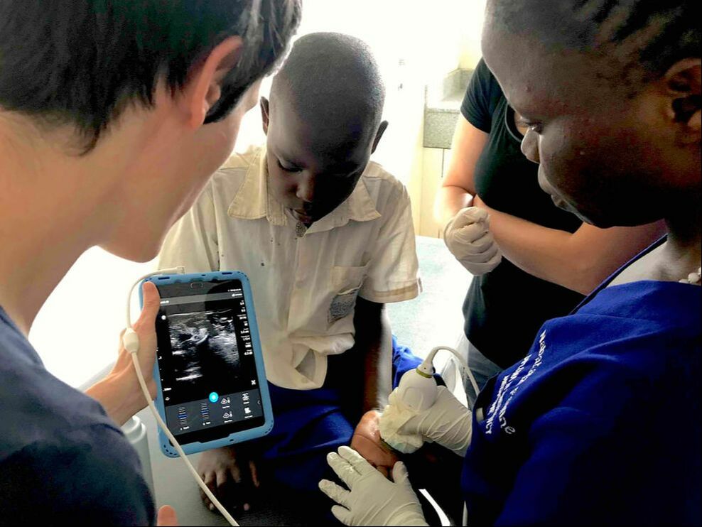
As our primary diagnostic tool, ultrasound can provide a fabulous amount of confirmatory and ancillary information. We first image the four quadrants of the abdomen, which in the upper abdomen demonstrates large, dilated loops of bowel with anterograde and retrograde peristalsis. This indicated the patient has a bowel obstruction.
In the lower abdomen, his bowel appears normal, which indicates the obstruction is higher than the bowel imaged. When we place the probe on the umbilicus we see intestine within it and adjacent free fluid. We have difficulty acquiring reliable color flow from the intestine.
We consult surgery immediately and the patient is taken to the operating theater. The surgeon successfully released the strangulated umbilical hernia. Even though we had feared intestinal necrosis and perforation, the surgeon found the bowel to be well perfused. The patient had an uneventful post-surgical course.
Ultrasound Machine Fundraiser for GEC
Thanks to a very generous gift from the Ellis family in honor of their parents, Dan and Barbara Ellis, we will be offering a 100% match on all ultrasound donations up to $10,000. Our goal in the next month is to raise $30,000 to fix one of our current ultrasound machines and to purchase two new ultrasound machines.
This Post Has 0 Comments